

Karyokinesis is the process of nuclear division during cell division.
- It is a crucial step in both mitosis and meiosis, ensuring that genetic material (DNA) is accurately distributed into the daughter cells.
- Without karyokinesis, proper cell division and inheritance of genetic information would not be possible.
- Division of a cell’s nucleus, during which the chromosomes are separated and distributed into two (or more) daughter nuclei.
Characteristics of Karyokinesis
- Karyokinesis is a process where the nucleus splits into two daughter nuclei during cell division.
- It is the first step of the M phase and occurs in both mitosis and meiosis.
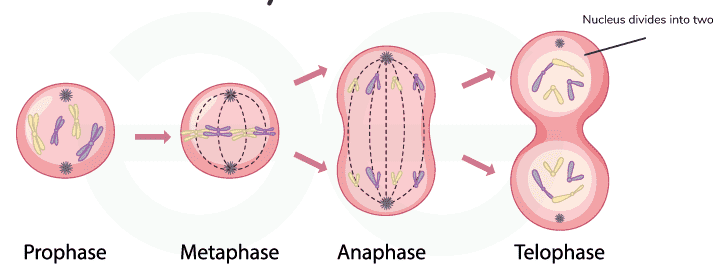
- During karyokinesis, DNA condenses into chromosomes, which are then properly aligned and equally distributed into two daughter nuclei.
- The process takes place in four main phases: prophase, metaphase, anaphase, and telophase.
- Karyokinesis is followed by cytokinesis, which involves the division of the cytoplasm and cell membrane.
- This process is essential for growth, repair, and regeneration in living organisms.
Phases of Karyokinesis
Karyokinesis is the process of nuclear division that occurs during cell division, specifically in mitosis or meiosis, and is particularly prominent in eukaryotic cells. It is the first step in the M phase of the cell cycle. It is followed by cytokinesis, which is the splitting of the cytoplasm and cell membrane.
Karyokinesis is divided into four phases: Prophase, Metaphase, Anaphase, and Telophase.
1. Prophase
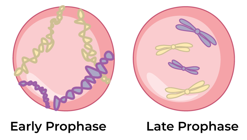
Prophase is the initial stage of both mitosis and meiosis. It is the longest phase and is divided into two phases:
Early Prophase
- It is the first stage of mitosis that follows G₂ phase (S phase is part of interphase but not directly before prophase)
- Chromatin fibres begin to condense into visible chromosomes.
- The mitotic spindle starts forming.
- Centrioles duplicate and move to the opposite poles of the cell.
Late Prophase (Prometaphase)
- The nuclear envelope completely breaks down.
- Spindle fibres attach to the kinetochores of chromosomes.
- Chromosomes begin to move toward the equatorial plane.
- Kinetochores are linked to spindle microtubules, enabling chromosome movement.
2. Metaphase
- It is the second stage of mitosis that follows prophase.
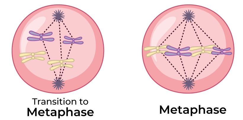
- Chromosomes align at the equatorial plane of the cell, forming the metaphase plate.
- The chromosomes are at their most condensed and coiled stage during metaphase.
- Spindle fibres from opposite poles attach to the kinetochore of each chromosome.
- Chromosomes are positioned for equal distribution to daughter cells in the upcoming anaphase.
3. Anaphase
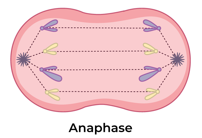
- Anaphase is the third mitosis phase, where chromosomes move to opposite poles.
- The centromeres split, allowing sister chromatids to separate.
- Kinetochore microtubules shorten.
- Non-kinetochore microtubules lengthen.
- Centrosomes are pushed far apart, and the cell elongates as spindle fibres continue to separate chromatids.
- This phase ensures that each daughter cell receives a complete set of chromosomes for genetic stability.
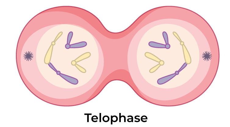
4. Telophase
- Telophase is the final stage of karyokinesis, followed by cytokinesis, dividing the cytoplasm.
- It starts when the replicated, paired chromosomes have been separated and pulled to opposite sides, or poles, of the cell.
- Chromosomes start to decondense into chromatin.
- The spindle apparatus gradually disappears.
- Nuclear envelopes re-form around each set of chromosomes.
- The nucleolus reappears in each daughter nucleus.
- Two distinct daughter nuclei are formed.
Karyokinesis in Meiosis
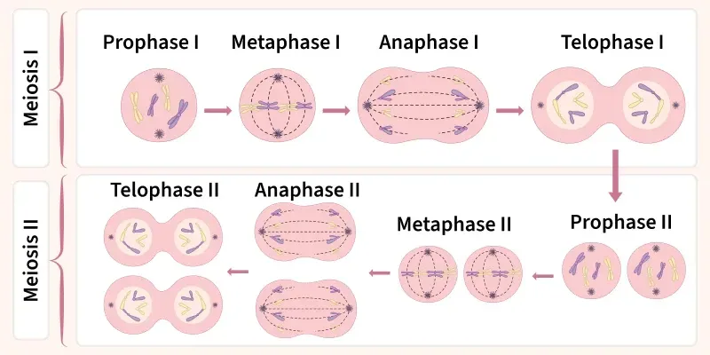
Meiosis consists of two successive cell divisions, known as meiosis I and meiosis II:
Meiosis I
- Prophase I: Chromosomes condense, and homologous chromosomes undergo genetic recombination (crossing over).
- Metaphase I: Homologous chromosomes align at the cell's equator.
- Anaphase I: Homologous chromosomes separate and move to opposite poles.
- Telophase I: Two haploid cells are formed, each with half the chromosome number of the original cell.
Meiosis II
Similar to mitosis, resulting in the formation of four non-identical haploid daughter cells.
- Prophase II: Chromosomes condense again, and spindle fibres form.
- Metaphase II: Chromosomes align at the equatorial plane.
- Anaphase II: Sister chromatids separate and move to opposite poles.
- Telophase II: Four non-identical haploid daughter nuclei are formed.
Significance of Karyokinesis
- It ensures the accurate distribution of genetic material, including chromosomes, to daughter cells during cell division.
- It helps in maintaining genetic stability and preserving the species-specific information encoded in the DNA.
- The process is fundamental for growth, tissue repair, and the maintenance of the organism's overall structure.
- It is important for living organisms because it ensures that cells can regenerate.
- It ensures that cells and tissues can mature, growth and development.
