

The mechanism of muscle contraction refers to the series of events through which a muscle fibre generates force and produces movement. Muscle contraction occurs when a nerve impulse stimulates a muscle fibre, leading to interactions between actin and myosin filaments within the muscle cells. The process involves the conversion of chemical energy stored in ATP into mechanical energy that results in muscle shortening and movement.
Structure of the Sarcomere
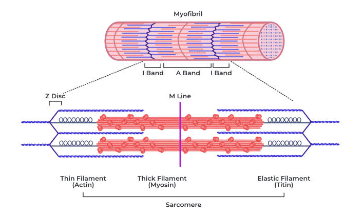
The sarcomere is the structural and functional unit of a myofibril and is responsible for muscle contraction. It is the region between two successive Z-lines or Z-discs. The precise arrangement of these filaments gives skeletal muscles their characteristic striated appearance. A sarcomere contains two main types of protein filaments:
- Thin filaments made primarily of actin.
- Thick filaments made primarily of myosin.
Bands and Regions of the Sarcomere
- A-band (Anisotropic Band): The A-band is the dark region of the sarcomere and corresponds to the entire length of the thick myosin filaments. Since it contains thick filaments throughout its length, it appears darker under a microscope.
- H-zone: The H-zone is the central region of the A-band where only thick myosin filaments are present. Thin actin filaments do not extend into this region when the muscle is relaxed.
- Zone of Overlap: The zone of overlap is the region where thin actin filaments overlap with thick myosin filaments. It is within this region that cross-bridges form during muscle contraction.
- I-band (Isotropic Band): The I-band is the light-coloured region containing only thin actin filaments. It is bisected by the Z-line.
- Z-line: The Z-line forms the boundary of each sarcomere and serves as the attachment point for the thin filaments.
Sliding Filament Theory
According to the sliding filament theory, muscle contraction occurs because the thin actin filaments slide inward toward the centre of the sarcomere over the thick myosin filaments. The filaments themselves do not shorten; instead, their degree of overlap increases. As a result:
- The Z-lines move closer together.
- The sarcomere becomes shorter.
- The H-zone decreases in size or disappears.
- The I-band becomes narrower.
- The A-band remains unchanged because the length of the myosin filament does not change.
Mechanism of Muscle Contraction
- Excitation of the Muscle Fibre: The process begins when a nerve impulse or action potential travels along a motor neuron and reaches the neuromuscular junction, also known as the motor end plate.
- Release of Acetylcholine: When the nerve impulse reaches the synaptic terminal, synaptic vesicles release the neurotransmitter acetylcholine into the synaptic cleft. Acetylcholine diffuses across the cleft and binds to specific receptors on the sarcolemma of the muscle fibre. This binding generates an action potential in the muscle membrane.
- Transmission Through T-Tubules: The action potential spreads rapidly across the sarcolemma and enters the muscle fibre through transverse tubules or T-tubules. These tubules carry the electrical signal deep into the muscle fibre, ensuring that all myofibrils contract simultaneously.
- Release of Calcium Ions: The action potential stimulates the sarcoplasmic reticulum, a specialized endoplasmic reticulum of muscle cells. In response, the sarcoplasmic reticulum releases calcium ions (Ca²⁺) into the sarcoplasm surrounding the myofibrils.
- Binding of Calcium to Troponin: The released calcium ions bind to the protein troponin present on the actin filaments. Under resting conditions, another protein called tropomyosin covers the myosin-binding sites on actin. When calcium binds to troponin, it causes a conformational change that moves tropomyosin away from these binding sites.
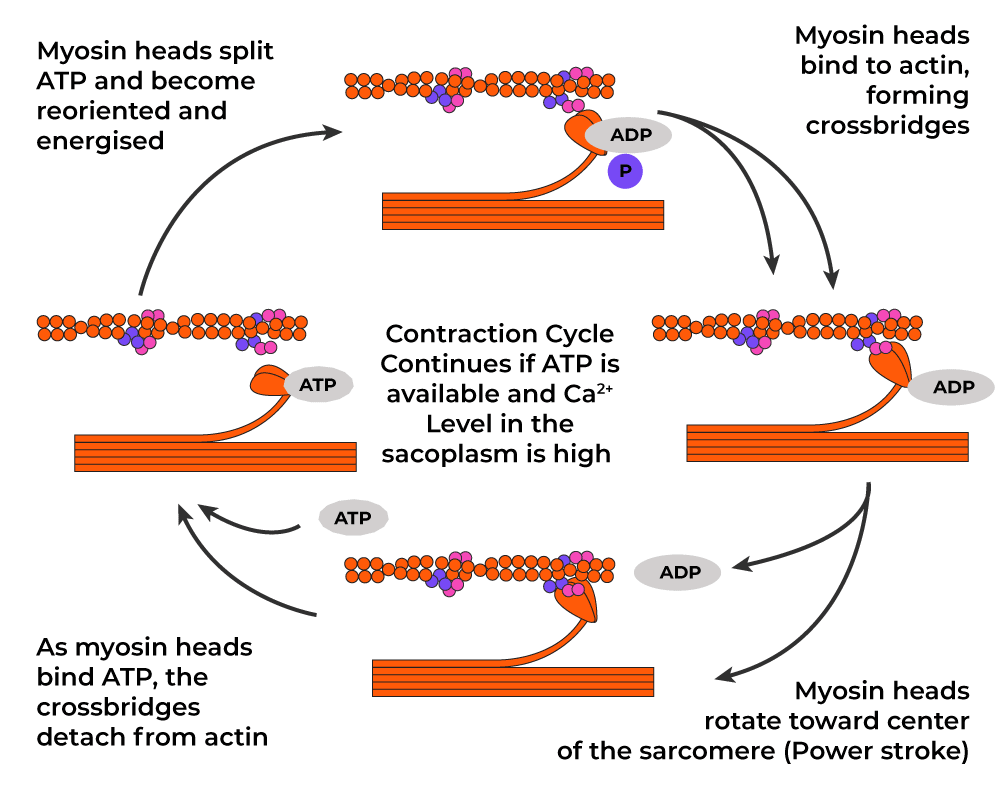
- Formation of Cross-Bridges: The myosin head contains stored energy obtained from the hydrolysis of ATP. The energized myosin head attaches to the exposed binding site on actin, forming a structure known as a cross-bridge.
- Power Stroke: Following cross-bridge formation, the myosin head pivots toward the center of the sarcomere. This movement is known as the power stroke. During the power stroke, the actin filament is pulled inward toward the center of the sarcomere. As millions of cross-bridges perform this action simultaneously, the muscle fibre contracts.
- Detachment of the Cross-Bridge: After the power stroke is completed, a new ATP molecule binds to the myosin head. The binding of ATP causes the myosin head to detach from the actin filament.
- Reactivation of the Myosin Head: The ATP molecule is hydrolysed into ADP and inorganic phosphate (Pi). The energy released re-cocks the myosin head into its high-energy position, preparing it for another cycle of cross-bridge formation. As long as calcium ions and ATP remain available, this cycle repeats continuously and muscle contraction continues.
Muscle Relaxation
Muscle relaxation begins when nerve stimulation ceases. The following events occur:
- The release of acetylcholine stops.
- Acetylcholine present in the synaptic cleft is rapidly broken down by the enzyme acetylcholinesterase.
- Calcium ions are actively pumped back into the sarcoplasmic reticulum using ATP.
- The concentration of calcium ions in the sarcoplasm decreases.
- Troponin returns to its original shape.
- Tropomyosin once again covers the myosin-binding sites on actin.
- Cross-bridge formation ceases.
- Actin filaments slide back to their resting position.
- The muscle fibre returns to its relaxed state.
Summary of Changes During Muscle Contraction
- Z-lines move closer together.
- Sarcomere length decreases.
- H-zone narrows or disappears.
- I-band becomes shorter.
- A-band remains unchanged.
- Overlap between actin and myosin filaments increases.
